Sunday, 29 March 2009

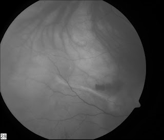
Odd submacular lesion with fibrosis

This right eye was initially treated with a haemorrhagic PED in early 2007, presenting with a vision of 6/24. Three Avastin injections were given at 6 weekly intervals, and things settled nicely with vision varying between 6/18 and 6/12 on subsequent visits. The last injection was September 2007.

Things remained stable till Feb 09, when she came back with a new complain of seeing a 'blob' in her central vision in her right eye.
Vision was still 6/18
Ant Seg - unremarkable, no cells in her vitreous.
This is her colour fundus photo:

Red Free pictures

She has a clear history of Flourescien allergy, so we couldn't do one.
IR photo:


Her early ICG is shown


Late ICG

Early ICG movie
Mid-phase ICG movie

Late ICG movie

Other eye had PDT some years ago for a fibrovascular PED and settled to a vision of 6/36, been stable for 5+ years.

Any idea about diagnosis? Management?

Wednesday, 25 March 2009

Another 25g Retinal detachment video

Another video showing proper technique to overcome 'weaknesses' of 25g technology.

A good chandelier light and extensive use of deep indentation, means that surgical goals can be achieved, even with relatively flexible instruments.

10th Antwerp VR course 2009

Just got back from Antwerp, excellent meeting as usual in it's 10th edition.

Big play on air tamponade only for macular hole with 3 day face down posturing. I've done a few caes like this with mixed results, anyone else tried this?

The gauge wars continue - 25, 23 or 20? Different people propunding the virtues of one system over the other. What is the outside diameter of a 20g Trocar cannula system for transconjunctival surgery? Must be 19 gauge, if 20g instruments go through the cannula? Will have to look this one up.....

Som

Sunday, 15 March 2009

Inducing a PVD

Usually it is easy to induce a posterior vitreous detachment, but some times the posterior hyaloid face can be very resistant to coming off...

Here is one such case, after trying all the usual manouvres which failed, I decided to 'stain' the vitreous with triamcinolone and then mechanically peel the posterior hyaloid face till I could develop the plane of separation. Once tha was achieved, engaging and pulling up the posterior hyaloid face with the ocutome was easy.


Humour - have a laugh for a change!

The Medical Profession Speaks Out On The Financial Bail-Out Package

The allergists voted to scratch it, and the dermatologists advised not to make any rash moves.

The gastroenterologists had sort of a gut feeling about it, but the neurologists thoughtthe administration had a lot of nerve, and the obstetricians felt they were all laboring under a misconception.

The ophthalmologists considered the idea shortsighted; the pathologists yelled,"Over my dead body!" while the pediatricians said, 'Oh, Grow up!'

The psychiatrists thought the whole idea was madness, the radiologists could see right through it,and the surgeons decided to wash their hands of the whole thing.

The internists thought it was a bitter pill to swallow,and the plastic surgeons said, "This puts a whole new face on the matter."

The podiatrists thought it was a step forward, but the urologists felt the scheme wouldn't hold water.

The anesthesiologists thought the whole idea was a gas; and the cardiologists didn't have the heart to say no.

In the end, the proctologists left the decision up to the assholes in Washington.

Wednesday, 11 March 2009

25g vitrectomy for retinal detachment

I was asked to review a paper for one of the journals on a large series (300+) of 25g transconjunctival vitrectomies. This brought back the arguments for and against this technique to the fore.

25G techniques have been blamed for the flexibility of instruments, the percieved inability to do peripheral work, and in the past for poor light. The light issue has long been sorted by modern sources such as the Photon. The flexibility of instruments, whilst real, can be overcome by changing technique. For instance when 'cleaning' a hole near the ora (very peripheral), traditionally one has the eye in a very tilted position to access the break. However, if you keep the eye flat - and use indentation to bring the area of interest to the vitrector, this problem is solved.

Here is a video clip of a round hole - very near the ora serrata being cleared of vitreous. Eye is flat, tndentation is used to bring the area of the hole to the cutter, thus avoiding the need to tilt the eye and providing a very controlled, very peripheral vitrectomy.

Monday, 9 March 2009

Laser - from where?

I've been doing a bit of reading about how lasers came to be realised as a tool for the multitude of applications it is used for.

As retinal specialists, we use lasers all the time - in clinics and in the operating theatre.

We've all read about the introduction of opthalmic lasers, but ever wondered where lasers originated...

The clue is Albert Einstein of e=mc2 fame. Besides the theory of relativity, he has many other great ideas to his name. Einstein predicted lasers in 1917!
http://www.aps.org/publications/apsnews/200508/history.cfm

Vitreous base shaving

Ensuring a near complete excision of vitreous base is made much easier with the use of triamcinolone staining. Visulaisation can be further enhanced by using the light pipe as an indentation instrument, as the light nicely transilluminates the kenalog crystals in the vitreous base.

Sunday, 8 March 2009

What am I reading?

Ever wondered about the competency of sutureless incisions for vitrectomy? Well the answer may be in this recent rticle in the American Journal of Ophthalmology.
It seems that a bevelled 25g incision is most competent of the sutureless incisions - however the sutured incisons are still the most secure.

Vitreo-macular traction

I used to find diagnosing a Vitreo-macular traction very difficult before the advent of routine OCT. OCT has really enhanced our understanding of the retinl pathology, and 3D rendering can show the pathology even better.

VMT shown clearly on OCT here:

I offerred this patient surgery rather than laser or intravitreal injections and he did very well.

A 3D rendering of VMT -


Drainage of SRF through the tear

Internal drainage of subretinal fluid through the retinal tear in a fresh retinal detachment using a 25g system. As the air comes in the retina flattens nicely. This is a clip from 5 years ago, when the 25g light sourcses were not very bright, but current light sources are both brighter and provide a wide field of illumination.

Follow Blog

Welcome to all the new visitors - If you are wondering how to keep track of the blog posts, why not click on the 'follow' button to the right.
If you are wondering waht this is - a good help file is here

Friday, 6 March 2009

Technical

Colleagues,

I have used two methods to post videos on this blog:
1. Upload to Blogger - these are the videos which appear witha an orange blogger icon in the lower right area
2. Microsoft Silverlight - maybe offers better quality?

For a comparison

See this post - where both formats are available

Retina Surgeon: Does a Wiess ring signify a complete or near complete PVD?

Which do you prefer? It is an important question, as a surgical discussion is best being video based, however image quality, accessebility - ease of download and viewing and similar usability factors are very important

Lets have your thoughts so we can develop the blog accordingly

Best wishes

Som

Completely sutureless combined phaco and pars plana vitrectomy

I'm a great fan of sutureless surgery, and have been using sutureless 25g technology for the majority of my vitrectomy procedures since 2003. Of course the later introduction of 23g & then 20g sutureless systems brought in other possibilities...but let's leave that story for another day.....

When doing combined phaco and vitrectomy surgery, even with a 2.85 mm phaco clear corneal incision, I have routinely put a stitch into the corneal incision before going to the post segment procedure, as otherwise, I would sometimes loose the AC during peripheral indentattion whilst completing the peripheral vitrectomy and internal search.

The introduction of sub 2mm phaco technology has solved this problem. Theoretically, the most secure corneal wound is square, and a 1.6-1.8mm wide corneal incision which is of a similar length gets to this theoretical ideal.

So now, the procedure is:
1. Place an inferotemporal 25G cannula in the pars plana and plug it.
2. Do a bimanual microincision phaco using 2x 1.6 mm incisions
3. Implant a microincision lens using wound assisted technique through a sub 2 mm incision
4. Place the infusion line into the preplaced 25g cannula
5. Place two superior 25g ports
6. Complete pars plana vitrectomy (and associated procedures)
7. Remove 25g cannulas
Voila...done

Here is a video of a combined phaco, vity for mac hole case:

Steps 1 to 5 are as above, then you can see the posterior hyaloid being detached and complete vitrectomy with vitreous base shave using indentation being done. After ILM peel a FAX (Fluid air exchange) is done which finishes with direct drainage from the hole - allowing the hole to close on the table- air is left in as tamponade.

Then 25g ports are removed - and in spite of agressive indentation and peripheral vitrectomy during the posterior segment procedure, the AC remains sealed, deep and well formed.

Thursday, 5 March 2009

can you push back Vit from ant to post segments effectively?

Following blunt trauma this girl presented with vitreous in the AC - formed vitreous - also a GRT (Giant Retinal Tear). At surgery, I thought that getting the vitreous out of the way in the AC - could be a good idea - so after instilling some Kenalog into AC to highlight Vit, I tried injecting into AC to push vit back into PC - did it work? - Watch the video and comment...

Sunday, 1 March 2009

Interactive discussion

Welcome to everyone who has joined over the last few days.

Please add comments to posted cases, and post your own cases so that the interaction is lively and meaningful.

If you have any problems posting do get in touch and I can help. Email link

Next steps in the oil in AC case

So having not succeeded in controlling his intraocular pressure medically and the iris still being up against the endothelium, there was little choice but to go back to the operation theatre... Making two paracentecis, the AC was reformed using a Rycroft cannula on BSS, an inferior PI was created - see video-


I resisted the temptation to remove oil from the AC, as in the past when I have tried to do so, the oil from the post segment keeps coming forward, and I didn't want to loose tamponade, as the patient was still in the first week of a Giant Retinal Tear repair. He postured face down for 24 hours after this, and the oil in the AC went back to the posterior segment, and his intraocular pressure remained normal and his anterior chamber remained deep. 8 weeks later, I removed his silicon oil via a pars plana approach and his retina remains attached - mission accomplished!

Question: Should I have made an inferior PI at his primary surgery? This is always done for an aphakic eye, but in phakic/pseudophakic/combined IOL vity sil oil cases I have not done this usually?

Thoughts?